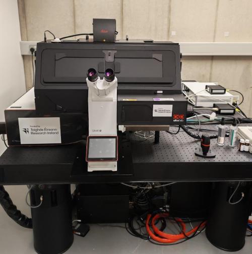
Leica Stellaris CRS 8 Confocal Microscope
Overview:
Confocal microscopy offers several advantages over conventional widefield optical microscopy, including the ability to control depth of field, elimination or reduction of background information away from the focal plane (that leads to image degradation), and the capability to collect serial optical sections from thick specimens. The basic key to the confocal approach is the use of spatial filtering techniques to eliminate out-of-focus light or glare in specimens whose thickness exceeds the immediate plane of focus.
The white light laser (WLL) of the Leica Stellaris SP8 system allows excitation across the range of 440 nm -790nm, tunable to match the spectral profile of the sample, and the simultaneous screening of 8 different spectral channels. Environmental control of the sample chamber enables, for example, live cell imaging.
The pulsed WLL technology also enables photon arrival time measurements (in a similar way to time correlated single photon counting), and therefore imaging based on fluorescence lifetime. The facility can be employed to contrast local microenvironments with emission profiles of different lifetimes, categorise fluorophores of multiple different emission lifetimes by gating (up to 16), and to determine fluorescence lifetimes and their distribution across a sample.
Leica Stellaris CRS 8 Confocal Microscope
The picosecond laser source opens up the possibilities for nonlinear optical based microscopic imaging techniques, including two photon fluorescence excitation (TPE), second harmonic generation (SHG), and Coherent Raman Scattering (CRS) microscopy, including coherent anti-Stokes (CARS), and stimulated Raman scattering (SRS).
TPE microscopy, typically performed in the near IR, provides increased penetration depth, particularly for tissue measurements.
SHG imaging is sensitive to non centrosymmetric structures at a molecular or morphological level, and in biological systems is commonly employed to probe structures of collagen, microtubules, and muscle myosin.
The techniques of CARS and SRS are third order nonlinear optical techniques based on Raman scattering, by which the frequency difference between pump (1032 nm) and probe (tuneable Optical Parametric Oscillator (720-980 nm)) lasers are tuned to exactly match that of a single vibrational frequency of a moiety of interest. The signal is enhanced by several orders of magnitude compared to incoherent Raman, and thus the spectroscopic signature of a sample can be mapped effectively at sampling times of microseconds per spot, while maintaining high signal to noise. CARS is typically used to rapidly map discrete frequencies, , whereas the full spectrum of a point or region of interest is measured by scanning the SRS responses over the full spectral range. Used in combination, the two techniques enable rapid, label fee, video rate chemical profiling of samples and processes.
Technical Specifications:
STELLARIS 8
- 405nm solid state laser
- WLL tunable from 440 nm to 790 nm, with 1nm precision.
- Simultaneous detection over 8 channels
- TauSense fluorescence lifetime imaging
CRS laser picoEmerald from APE
- Pump laser: Optical Parametric Oscillator (OPO) – tunable from 720 nm - 980 nm.
- Stokes laser: Fiber laser, fixed at 1032 nm, frequency doubled to pump the OPO.
- 20-MHz EOM intensity modulation of the Pump laser for SRS.
- CRS optimised Beam Routing.
- Dedicated IR objectives for optimised CRS signal generation and detection.
- Accessible range of vibrational frequencies for SRS: 4190 – 507 cm-1, covering the entire high-wavenumber, cell-silent, and fingerprint regions.
- Accessible range of vibrational frequencies for CARS: 4190 – 1200 cm-1.
- Switch between Forward SRS and Forward-CARS detection. Simultaneous detection of Epi-CARS signals.
- Truly multi-modal nonlinear optical microscopy with 2-Channel Detector Option: Simultaneous detection of CARS and Second-Harmonic Generation (SHG)/2-Photon Fluorescence signals (available for Forward and Epi detection).